9
Weak scattered light induced by laser beam entered into the anterior chamber is detected and used for measurements.
It is known that intensity of the scattered light is proportional to the protein level contained in aqueous humor of the anterior
chamber. However, intensity of the scattered light may vary if there is a difference in the protein composition between aqueous
humor samples at the same protein concentration.
We refer to this scattered light intensity as “flare value" and indicates the value using photon count per millisecond in this device.
✽ Photon count is the number of pulses output from photomultiplier when scattered light is detected. This value may be converted
into an albumin level. Bovine albumin solution at 100mg/dl equals to 13 Photon counts per millisecond.
■ Details ■
Measurements
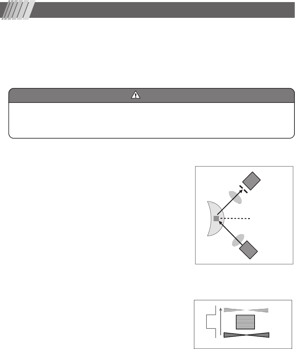
Optical system is composed of a laser beam emitter and a photoreceiver
positioned at a orthogonal to the axis of the beam. The scanning laser beam
emitted through a condensing lens is focused at the anterior chamber or
target point. Scattered light from the anterior chamber goes through a
photoreceiver lens and comes into a focus at a photoreceiver mask. The
photoreceiver mask has an important role to create a reading window within
aqueous humor of the anterior chamber. Scattered light coming through the
mask reach to a photoreceiver element (or a photomultiplier tube) where it
undergoes a photo-electro conversion process. Then, the collected data is
analyzed at the analyzer unit to determine a flare value. Results are shown
in the display.
Details of flare reading
An area including the Measurement window is scanned with laser beam. As
a result, a waveform shown in Fig. 1-2 is obtained. Background Signal 1
(BG1) obtained when laser beam is located below the a Measurement win-
dow and Background Signal 2 (BG2) obtained when laser beam is located
above the a Measurement window are scattered light noise from intraocular
tissue, while Flare Signal (SIG) is a sum of scattered light from protein and
scattered light noise from intraocular tissue.
Therefore, intensity of the scattered light caused by the protein concentration
in aqueous humor of the anterior chamber is calculated using the formula:
SIG - (BG1 + BG2)/2.
A result obtained using this formula is called “flare value” and represented
as photon count per millisecond.
CautionCaution
1. Principle of operation
BG1
SIG
BG2
Figure 1-1
Figure 1-2
Photoreceiver
element
Photoreceiver
mask
Anterior
chamber
Scanning laser
Measurement
window
Laser scanning light
Measurement
window
Photoreceiver lens
Condensing lens
Note that some factors including circadian rhythm, age, mydriasis, and drug may affect flare values. Measurement must be taken
carefully taking any of these factor in account. The accuracy of the reading may be affected by disorders shown below:
✽ Intensive lens clouding, corneal edema, corneal opacity, the anterior chamber with an artificial lens implanted, shallow anterior
chamber, and achromatic eye.


